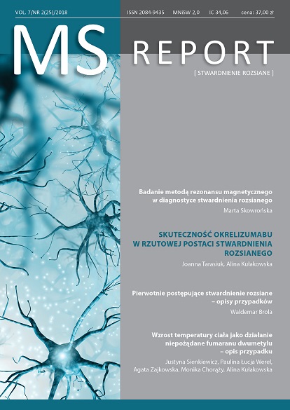
Magnetic resonance imaging in multiple sclerosis diagnosis Review article
Main Article Content
Abstract
According to recent recommendations for diagnosis of multiple sclerosis in patient with clinically isolated syndrome:
- dissemination in space can be demonstrated in magnetic resonance by 1 or more T2-hyperintense lesions that are characteristic of multiple sclerosis in 2 or more of 4 areas of the central nervous system: periventricular, cortical or juxtacortical, infratentorial brain regions, and the spinal cord
- dissemination in time can be demonstrated in magnetic resonance by the simultaneous presence of gadolinium-enhancing and non-enhancing lesions at any time or new T2-hyperintense or gadolinium-enhancing lesion on follow-up magnetic resonance imaging, with reference to a baseline scan, irrespective of the timing of the baseline magnetic resonance imaging.
No distinction between symptomatic and asymptomatic magnetic resonance imaging lesions is required.
Article Details
Issue
Section
Articles
Copyright © by Medical Education. All rights reserved.
References
1. McDonald W.I., Compston A., Edan G. et al.: Recommended diagnostic criteria for multiple sclerosis: guidelines from the International Panel on the diagnosis of multiple sclerosis. Ann. Neurol. 2001; 50(1): 121-127.
2. Polman C.H., Reingold S.C., Edan G. et al.: Diagnostic criteria for multiple sclerosis: 2005 revisions to the “McDonald Criteria”. Ann. Neurol. 2005; 58(6): 840-846.
3. Polman C.H., Reingold S.C., Banwell B. et al.: Diagnostic criteria for multiple sclerosis: 2010 Revisions to the McDonald criteria. Ann. Neurol. 2011; 69(2): 292-302.
4. Thompson A.J., Banwell B.L., Barkhof F. et al.: Diagnosis of multiple sclerosis: 2017 revisions of the McDonald criteria. The Lancet Neurology 2018; 17(2): 162-173.
5. Filippi M., Rocca M.A., Ciccarelli O. et al.; on behalf of the MAGNIMS Study Group: MRI criteria for the diagnosis of multiple sclerosis: MAGNIMS Consensus Guidelines. Lancet Neurol. 2016; 15(3): 292-303.
6. Hemond C.C., Bakshi R.: Magnetic resonance imaging in multiple sclerosis. Cold Spring Harb. Perspect. Med. DOI: 10.1101/cshperspect.a028969.
7. Absinta M., Rocca M.A., Colombo B. et al.: Patients with migraine do not have MRI-visible cortical lesions. J. Neurol. 2012; 259: 2695-2698.
8. Kim S.S., Richman D.P., Johnson W.O. et al.: Limited utility of current MRI criteria for distinguishing multiple sclerosis from common mimickers: primary and secondary CNS vasculitis, lupus and Sjogren’s syndrome. Mult. Scler. 2014; 20(1): 57-63.
9. Seewann A., Vrenken H., Kooi E.J. et al.: Imaging the tip of the iceberg: visualization of cortical lesions in multiple sclerosis. Mult. Scler. 2011; 17: 1202-1210.
10. Seewann A., Kooi E.J., Roosendaal S.D. et al.: Postmortem verification of MS cortical lesion detection with 3D DIR. Neurology 2012; 78(5): 302-308.
11. Calabrese M., Oh M.S., Favaretto A. et al.: No MRI evidence of cortical lesions in neuromyelitis optica. Neurology 2012; 79(16): 1671-1676.
12. Brownlee W.J., Hardy T.A., Fazekas F., Miller D.H.: Multiple Sclerosis 1. Diagnosis of multiple sclerosis: progress and challenges. Lancet 2016; 389: 1336-1346.
13. Kearney H., Miller D.H., Ciccarelli O.: Spinal cord MRI in multiple sclerosis – diagnostic, prognostic and clinical value. Nat. Rev. Neurol. 2015; 11: 327-338.
14. Masdeu J.C., Quinto C., Olivera C. et al.: Open-ring imaging sign: Highly specific for atypical brain demyelination. Neurology 2000; 54: 1427-1433.
15. Minneboo A., Uitdehaag B.M.J., Ader H.J. et al.: Patterns of enhancing lesion evolution in multiple sclerosis are uniform within patients. Neurology 2005; 65: 56-61.
16. Molyneux P.D., Filippi M., Barkhof F. et al.: Correlations between monthly enhanced MRI lesion rate and changes in T2 lesion volume in multiple sclerosis. Ann. Neurol. 1998; 43: 332-339.
17. Kappos L., Moeri D., Radue E.W. et al.: Predictive value of gadolinium-enhanced magnetic resonance imaging for relapse rate and changes in disability or impairment in multiple sclerosis: A meta-analysis. Lancet 1999; 353: 964-969.
18. van Walderveen M.A., Kamphorst W., Scheltens P. et al.: Histopathologic correlate of hypointense lesions on T1-weighted spin-echo MRI in multiple sclerosis. Neurology 1998; 50(5): 1282-1288.
19. Sahraian M.A., Radue E.W., Haller S., Kappos L.: Black holes in multiple sclerosis: Definition, evolution, and clinical correlations. Acta Neurol. Scand. 2010; 122: 1-8.
20. Mitjana R., Tintore M., Rocca M.A. et al.: Diagnostic value of brain chronic black holes in T1-weighted MR images in clinically isolated syndromes. Mult. Scler. J. 2014; 20: 1471-1477.
21. Okuda D.T., Mowry E.M., Beheshtian A. et al.: Incidental MRI anomalies suggestive of multiple sclerosis: the radiologically isolated syndrome. Neurology 2009; 72(9): 800-805.
22. De Stefano N., Giorgio A., Tintore M. et al.; on behalf of the MAGNIMS study group: Radiologically isolated syndrome or subclinical multiple sclerosis: MAGNIMS consensus recommendations. Mult. Scler. 2018; 24(2): 214-221.
23. Okuda D.T., Mowry E.M., Cree B.A. et al.: Asymptomatic spinal cord lesions predict disease progression in radiologically isolated syndrome. Neurology 2011; 76(8): 686-692.
24. Lebrun C., Bensa C., Debouverie M. et al.: Association between clinical conversion to multiple sclerosis in radiologically isolated syndrome and magnetic resonance imaging, cerebrospinal fluid, and visual evoked potential: Follow-up of 70 patients. Arch. Neurol. 2009; 66(7): 841-846.

