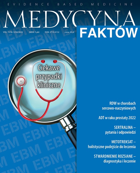
Red cell distribution width as a prognostic indicator in cardiovascular diseases Review article
Main Article Content
Abstract
Red cell distribution width is a reflection of the erythrocyte size distribution and is, therefore, a reliable and meaningful indicator of anisocytosis commonly used in the differential diagnosis of micro, macro, and normocytic anemia. Based on recent scientific reports in recent decades, it has been observed that the level of red cell distribution width significantly reflects a worsening prognosis and is associated with the presence of complications in patients suffering from cardiovascular diseases.
This article aims to discuss the most important scientific reports on the correlation of red cell distribution width values with cardiovascular incidents and mortality in order to assess its usefulness as a cheap and readily available marker in clinical practice. Using the available data, it seems reasonable to claim that abnormal red cell distribution width values should induce further expansion of the diagnosis of anemia and cardiovascular disease.
Article Details
Copyright © by Medical Education. All rights reserved.
References
2. Auerbach M, Adamson JW. How we diagnose and treat iron deficiency anemia. Am J Hematol. 2016; 91(1): 31-8. http://doi.org/10.1002/ajh.24201.
3. Desalegn Wolide A, Mossie A, Gedefaw L. Nutritional iron deficiency anemia: magnitude and its predictors among school age children, southwest Ethiopia: a community based cross-sectional study. PLoS One. 2014; 9(12): e114059. http://doi.org/10.1371/journal.pone.0114059.
4. Diagnostic value of the mean corpuscular volume in the detection of vitamin B12 deficiency. Scand J Clin Lab Invest. 2000; 60(1): 9-18. http://doi.org/10.1080/00365510050184994.
5. Dugdale AE. Predicting iron and folate deficiency anaemias from standard blood testing: the mechanism and implications for clinical medicine and public health in developing countries. Theor Biol Med Model. 2006; 3: 34. http://doi.org/10.1186/1742-4682-3-34.
6. Mahmood NA, Mathew J, Kang B et al. Broadening of the red blood cell distribution width is associated with increased severity of illness in patients with sepsis. Int J Crit Illn Inj Sci. 2014; 4(4): 278-82. http://doi.org/10.4103/2229-5151.147518.
7. Maruyama S, Hirayama C, Yamamoto S et al. Red blood cell status in alcoholic and non-alcoholic liver disease. J Lab Clin Med. 2001; 138(5): 332-7. http://doi.org/10.1067/mlc.2001.119106.
8. Nagano T, Toyoda T, Tanabe H et al. Clinical features of hematological disorders caused by copper deficiency during long term enteral nutrition. Intern Med. 2005; 44(6): 554-9. http://doi.org/10.2169/internalmedicine.44.554.
9. Baker RD, Greer FR; Committee on Nutrition American Academy of Pediatrics. Diagnosis and prevention of iron deficiency and iron-deficiency anemia in infants and young children (0-3 years of age). Pediatrics. 2010; 126(5): 1040-50. http://doi.org/10.1542/peds.2010-2576.
10. Tussing-Humphreys L, Pusatcioglu C, Nemeth E et al. Rethinking iron regulation and assessment in iron deficiency, anemia of chronic disease, and obesity: introducing hepcidin. J Acad Nutr Diet. 2012; 112(3): 391-400. http://doi.org/10.1016/j.jada.2.
11. Bottomley SS, Fleming MD. Sideroblastic anemia: diagnosis and management. Hematol Oncol Clin North Am. 2014; 28(4): 653-70. http://doi.org/10.1016/j.hoc.2014.04.008.
12. Francis J, Sheridan D, Samanta A et al. Iron deficiency anaemia in chronic inflammatory rheumatic diseases: low mean cell haemoglobin is a better marker than low mean cell volume. Ann Rheum Dis. 2005; 64(5): 787-8. http://doi.org/10.1136/ard.2004.025890.
13. Aslinia F, Mazza JJ, Yale SH. Megaloblastic anemia and other causes of macrocytosis. Clin Med Res. 2006; 4(3): 236-41. http://doi.org/10.3121/cmr.4.3.236. Erratum in: Clin Med Res. 2006; 4(4): 342.
14. Savage DG, Ogundipe A, Allen RH et al. Etiology and diagnostic evaluation of macrocytosis. Am J Med Sci. 2000; 319(6): 343-52. http://doi.org/10.1097/00000441-200006000-00001.
15. Dorgalaleh A, Mahmoodi M, Varmaghani B et al. Effect of thyroid dysfunctions on blood cell count and red blood cell indice. Iran J Ped Hematol Oncol. 2013; 3(2): 73-7.
16. Conway AM, Vora AJ, Hinchliffe RF. The clinical relevance of an isolated increase in the number of circulating hyperchromic red blood cells. J Clin Pathol. 2002; 55(11): 841-4. http://doi.org/10.1136/jcp.55.11.841.
17. Wu TT, Zheng YY, Hou XG et al. Red blood cell distribution width as long-term prognostic markers in patients with coronary artery disease undergoing percutaneous coronary intervention. Lipids Health Dis. 2019; 18: 140. http://doi.org/10.1186/s12944-019-1082-8.
18. Fava C, Cattazzo F, Hu ZD et al. The role of red blood cell distribution width (RDW) in cardiovascular risk assessment: useful or hype? Ann Transl Med. 2019; 7(20): 581. http://doi.org/10.21037/atm.2019.09.58.
19. Bujak K, Wasilewski J, Osadnik T et al. The Prognostic Role of Red Blood Cell Distribution Width in Coronary Artery Disease: A Review of the Pathophysiology. Dis Markers. 2015; 2015: 824624. http://doi.org/10.1155/2015/824624.
20. Alcaíno H, Pozo J, Pavez M et al. Ancho de distribución eritrocitaria como potencial biomarcador clínico en enfermedades cardiovasculares. Revista médica de Chile. 2016; 144(5): 634-42. https://dx.doi.org/10.4067/S0034-98872016000500012.
21. Baggen VJM, van den Bosch AE, van Kimmenade RR et al. Red cell distribution width in adults with congenital heart disease: A worldwide available and low-cost predictor of cardiovascular events. Int J Cardiol. 2018; 260: 60-5. http://doi.org/10.1016/j.ijcard.2018.02.118 .
22. Danese E, Lippi G, Montagnana M. Red blood cell distribution width and cardiovascular diseases. J Thorac Dis. 2015; 7(10): E402-11. http://doi.org/10.3978/j.issn.2072-1439.2015.10.04.
23. Abrahan LL 4th, Ramos JDA, Cunanan EL et al. Red Cell Distribution Width and Mortality in Patients With Acute Coronary Syndrome: A Meta-Analysis on Prognosis. Cardiol Res. 2018; 9(3): 144-52. http://doi.org/10.14740/cr732w.
24. Isik T, Ayhan E, Kurt M et al. Is red cell distribution width a marker for the presence and poor prognosis of cardiovascular disease? Eurasian J Med. 2012; 44(3): 169-71. http://doi.org/10.5152/eajm.2012.39.
25. Pan J, Borné Y, Gonçalves I et al. Associations of Red Cell Distribution Width With Coronary Artery Calcium in the General Population. Angiology. 2021: 33197211052124. http://doi.org/10.1177/00033197211052124.
26. Gili M, Ramírez G, Béjar L et al. Cocaine use disorders and acute myocardial infarction, excess length of hospital stay and overexpenditure. Rev Esp Cardiol (Engl Ed). 2014; 67(7): 545-51. http://doi.org/10.1016/j.rec.2013.11.010.
27. Lippi G, Turcato G, Cervellin G et al. Red blood cell distribution width in heart failure: A narrative review. World J Cardiol. 2018; 10(2): 6-14. http://doi.org/10.4330/wjc.v10.i2.6.
28. Dugdale AE, Badrick T. Red blood cell distribution width (RDW) – a mechanism for normal variation and changes in pathological states. J Lab Precis Med. 2018: n. pag.
29. He W, Jia J, Chen J et al. Comparison of prognostic value of red cell distribution width and NT-proBNP for short-term clinical outcomes in acute heart failure patients. Int Heart J. 2014; 55(1): 58-64. http://doi.org/10.1536/ihj.13-172.
30. Abdullah HR, Sim YE, Sim YT et al. Preoperative Red Cell Distribution Width and 30-day mortality in older patients undergoing non-cardiac surgery: a retrospective cohort observational study. Sci Rep. 2018; 8: 6226. http://doi.org/10.1038/s41598-018-24556-z.
31. Vizzardi E, Sciatti E, Bonadei I et al. Red cell distribution width and chronic heart failure: prognostic role beyond echocardiographic parameters. Monaldi Arch Chest Dis. 2016; 84(1-2): 59. http://doi.org/10.4081/monaldi.2015.59.
32. Salvatori M, Formiga F, Moreno-Gónzalez R et al. Red blood cell distribution width as a prognostic factor of mortality in elderly patients firstly hospitalized due to heart failure. Kardiol Pol. 2019; 77(6): 632-8. http://doi.org/10.33963/KP.14818.
33. Zhang Y, Wang Y, Kang JS et al. Differences in the predictive value of red cell distribution width for the mortality of patients with heart failure due to various heart diseases. J Geriatr Cardiol. 2015; 12(6): 647-54. http://doi.org/10.11909/j.issn.1671-5411.2015.06.001.
34. Kaya A, Isik T, Kaya Y et al. Relationship between red cell distribution width and stroke in patients with stable chronic heart failure: a propensity score matching analysis. Clin Appl Thromb Hemost. 2015; 21(2): 160-5. http://doi.org/10.1177/1076029613493658.
35. Altaf A, Khan MA, Alam A et al. Correlation of red cell distribution width with inflammatory markers and its prognostic value in patients with diabetes and coronary artery disease. Clin Diabetol. 2020; 9(3): 174-8. http://doi.org/10.5603/dk.2020.0017.
36. Tonelli M, Sacks F, Arnold M et al.; for the Cholesterol and Recurrent Events (CARE) Trial Investigators. Relation Between Red Blood Cell Distribution Width and Cardiovascular Event Rate in People With Coronary Disease. Circulation. 2008; 117(2): 163-8. http://doi.org/10.1161/CIRCULATIONAHA.107.727545.

