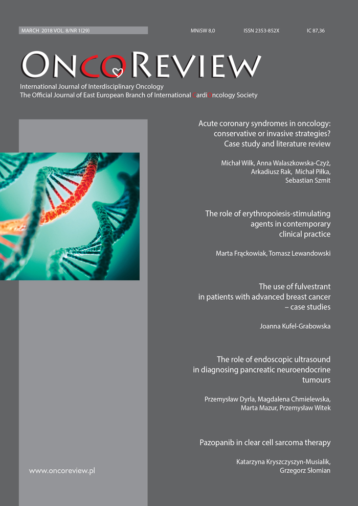
Znaczenie endosonografii w diagnostyce guzów neuroendokrynnych trzustki Review article
##plugins.themes.bootstrap3.article.main##
Abstrakt
Guzy trzustki stanowią jedno z największych wyzwań w diagnostyce obrazowej nowotworów układu pokarmowego. Guzy wywodzące się z wysp trzustkowych są drugą po gruczolakorakach – zdecydowanie rzadszą – grupą nowotworów trzustki. Nowotwory neuroendokrynne (NET, neuroendocrine tumors) charakteryzują się odmiennymi objawami i tempem przebiegu procesu rozrostowego, a tym samym wymagają innego podejścia diagnostycznego i terapeutycznego niż gruczolakoraki. Endosonografia (EUS, endoscopic ultrasound) wydaje się niezbędnym elementem w procesie diagnostycznym NET trzustki. Odznacza się także wysoką czułością i swoistością w ich wykrywaniu. Prawidłowy wynik EUS w praktyce wyklucza obecność guza trzustki. W przypadku zaś potwierdzonej obecności guza pozwala na dokładne określenie stosunków anatomicznych i ocenę zaawansowania procesu rozrostowego. Ogromną zaletą EUS jest możliwość wykonania biopsji i ocena cytologiczna/histopatologiczna pobranego materiału. Zastosowanie kontrastu i dodatkowych komputerowych metod analizy obrazu zwiększa trafność diagnostyczną endosonografii. W artykule przedstawiono aktualne poglądy na temat zastosowania EUS w diagnostyce nowotworów trzustki ze szczególnym uwzględnieniem nowotworów neuroendokrynnych.
Pobrania
##plugins.generic.paperbuzz.metrics##
##plugins.themes.bootstrap3.article.details##

Utwór dostępny jest na licencji Creative Commons Uznanie autorstwa – Użycie niekomercyjne 4.0 Międzynarodowe.
Copyright: © Medical Education sp. z o.o. This is an Open Access article distributed under the terms of the Attribution-NonCommercial 4.0 International (CC BY-NC 4.0). License (https://creativecommons.org/licenses/by-nc/4.0/), allowing third parties to copy and redistribute the material in any medium or format and to remix, transform, and build upon the material, provided the original work is properly cited and states its license.
Address reprint requests to: Medical Education, Marcin Kuźma (marcin.kuzma@mededu.pl)
Bibliografia
2. Wojciechowska U, Didkowska J, Zatoński W. Nowotwory złośliwe w Polsce w 2012 roku. Ministerstwo Zdrowia, Warszawa 2014.
3. Theoharis C. Mast cells and pancreatic cancer. N Engl J Med 2008; 358: 1860-1861.
4. Prokop M. Multidetector spiral computed tomography. Medipage, Warszawa 2007: 508-523.
5. Cascinu S, Falconi M, Valentini V et al. Pancreatic cancer: ESMO Clinical Practice Guidelines for diagnosis, treatment and follow-up. Ann Oncol 2010; 21: 55-58.
6. Seufferlein T, Bachet JB, Van Cutsem E et al. Pancreatic adenocarcinoma: ESMO-ESDO Clinical Practice Guidelines for diagnosis, treatment and follow-up. Ann Oncol 2012; 23(suppl 7): vii33-40.
7. Chari ST, Kelly K, Hollingsworth MA et al. Early detection of sporadic pancreatic cancer: summative review. Pancreas 2015; 44: 693-712.
8. Davis SL, Brooke RJ, Kamaya A. Islet-cell tumors of the pancreas: spectrum of MDCT findings. A pictorial essay. Applied Radiology 2009; 38: 11.
9. Kos-Kudła B, Blicharz-Dorniak J, Handkiewicz-Junak D et al. Diagnostic and therapeutic guidelines for gastro entero-pancreatic neuroendocrine neoplasms (recommended by the Polish Network of Neuroendocrine Tumours). Endokrynol Pol 2017; 68(2): 79 110; 169-197.
10. ENETS Consensus Guidelines Update for the Management of Patients with Functional Pancreatic Neuroendocrine Tumors and Non-Functional Pancreatic Neuroendocrine Tumors. Neuroendocrinology 2016; 103: 153-171.
11. Trofimiuk-Müldner M, Lewkowicz E, Wysocka K et al. Epidemiology of gastroenteropancreatic neuroendocrine neoplasms in Krakow and Krakow district in 2007-2011. Endokrynol Pol 2017; 68(1): 42-46.
12. Dimagno EP, Regan PT, Clain JE et al. Human endoscopic ultrasonography. Gastroenterology 1982; 83: 824-829.
13. Yusoff IF, Mendelson RM, Edmunds SE et al. Preoperative assessment of pancreatic malignancy using endoscopic ultrasound. Abdom Imaging 2003; 28: 556-562.
14. Kahl S, Glasbrenner B, Zimmermann S et al. Endoscopic ultrasound in pancreatic diseases. Dig Dis 2002; 20: 120-126.
15. DeWitt J, Devereaux B, Chriswell M et al. Comparison of endoscopic ultrasonography and multidetector computed tomography for detecting and staging pancreatic cancer. Ann Intern Med 2004; 141: 753-763.
16. Goldberg J, Rosenblat J, Khatri G et al. Complementary roles of CT and endoscopic ultrasound in evaluating a pancreatic mass. AJR Am J Roentgenol 2010; 194(4): 984-992.
17. Ito T, Hijioka S, Masui T et al. Advances in the diagnosis and treatment of pancreatic neuroendocrine neoplasms in Japan. J Gastroenterol 2017; 52(1): 9-18.
18. Gouya H, Vignaux O, Augui J et al. CT, endoscopic sonography, and a combined protocol for preoperative evaluation of pancreatic insulinomas. AJR Am J Roentgenol 2003; 181(4): 987-992.
19. Zimmer T, Scherübl H, Faiss S et al. Endoscopic ultrasonography of neuroendocrine tumours. Digestion 2000; 62(suppl 1): 45-50.
20. Anderson MA, Carpenter S, Thompson NW et al. Endoscopic ultrasound is highly accurate and directs management in patients with neuroendocrine tumors of the pancreas. Am J Gastroenterol 2000; 95(9): 2271-2277.
21. Manta R, Nardi E, Pagano N et al. Pre-operative Diagnosis of Pancreatic Neuroendocrine Tumors with Endoscopic Ultrasonography and Computed Tomography in a Large Series. J Gastrointestin Liver Dis 2016; 25(3): 317-321.
22. Gonçalves B, Soares J, Bastos P. Endoscopic Ultrasound in the Diagnosis and Staging of Pancreatic Cancer. GE Port. J Gastroenterol 2015; 22(4): 161-171.
23. Sugimoto M, Takagi T, Hikichi T et al. Efficacy of endoscopic ultrasonography-guided fine needle aspiration for pancreatic neuroendocrine tumor grading. World J Gastroenterol 2015; 21(26): 8118-8124.
24. Newman NA, Lennon AM, Edil BH et al. Preoperative endoscopic tattooing of pancreatic body and tail lesions decreases operative time for laparoscopic distal pancreatectomy. Surgery 2010; 148(2): 371-377.
25. Dyrla P, Gil J, Florek M et al. Elastography in pancreatic solid tumours diagnosis. Prz Gastroenterol 2015; 10(1): 41-46.
26. Giovannini M, Hookey LC, Bories E et al. Endoscopic ultrasound elastography: the first step towards virtual biopsy? Preliminary results in 49 patients. Endoscopy 2006; 38: 344-348.
27. Hocke M, Schulze E, Gottschalk P et al. Contrast-enhanced endoscopic ultrasound in discrimination between focal pancreatitis and pancreatic cancer. World J Gastroenterol 2006; 12: 246-250.
28. Saftoiu A, Vilmann P, Gorunescu F et al. Neural network analysis of dynamic sequences of EUS elastography used for the differential diagnosis of chronic pancreatitis and pancreatic cancer. Gastrointest Endosc 2008; 68: 1086-1094.

