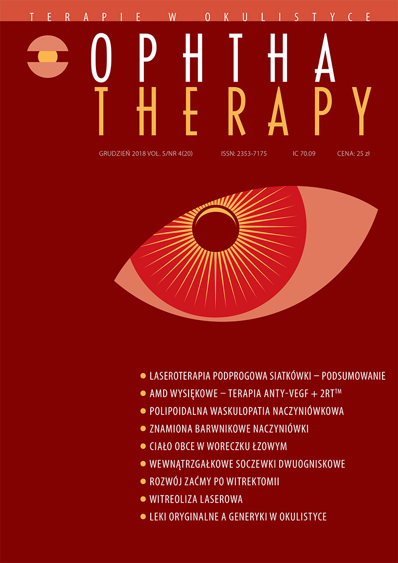
Diagnosis and management of polypoidal choroidal vasculopathy
Main Article Content
Abstract
Polypoidal choroidal vasculopathy is considered to be one of the subtypes of the neovascular age-related macular degeneration, especially frequent in Asians. The disease is clinically manifested by the recurrent serosanguineous pigment epithelial detachment and polypoid reddish-orange nodules visible in the posterior segment of the globe. The association of polypoidal choroidal vasculopathy with the spectrum of age-related macular degeneration raises some controversies due to the relatively rare occurrence in its case of several characteristic findings such as drusen, pigment changes or RPE atrophy. Choosing the optimal therapeutic option for each patient the individual characteristics of the patient's disease and the availability of diagnostic and therapeutic tools should be considered.
Downloads
Article Details

This work is licensed under a Creative Commons Attribution-NonCommercial-NoDerivatives 4.0 International License.
Copyright: © Medical Education sp. z o.o. License allowing third parties to copy and redistribute the material in any medium or format and to remix, transform, and build upon the material, provided the original work is properly cited and states its license.
Address reprint requests to: Medical Education, Marcin Kuźma (marcin.kuzma@mededu.pl)
References
2. Ferris FL III, Fine SL, Hyman L. Age-related maculardegeneration and blindness due to neovascular maculopathy. Arch Ophthalmol. 1984; 102: 1640-2.
3. Green WR, Enger C. Age-related macular degeneration histopathologic studies – the 1992 Zimmerman, Lorenz, E Lecture. Ophthalmology. 1993; 100: 1519-35.
4. Freund KB, Zweifel SA, Engelbert M. Do we need a new classification for choroidal neovascularization in age related macular degeneration? Retina. 2010; 30: 1333‐49.
5. Yannuzzi LA, Sorenson J, Spaide RF et al. Idiopathic polypoidal choroidal vasculopathy (IPCV). Retina. 1990; 10(1): 1-8.
6. Uyama M, Matsubara T, Fukushima I et al. Idiopathic polypoidal choroidal vasculopathy in Japanese patients. Arch Ophthalmol. 1999; 117: 1035-42.
7. Imamura Y, Engelbert M, Iida T et al. Polypoidal choroidal vasculopathy: a review. Surv Ophthalmol. 2010; 55(6): 501-15.
8. Lafaut BA, Leys AM, Snyers B et al. Polypoidal choroidal vasculopathy in Caucasians. Graefes Arch Clin Exp Ophthalmol. 2000; 238: 752-9.
9. Ma L, Li Z, Liu K et al. Association of genetic variants with polypoidal choroidal vasculopathy: a systematic review and updated meta-analysis. Ophthalmology. 2015; 122(9): 1854-65.
10. Woo SJ, Ahn J, Morrison MA et al. Analysis of genetic and environmental risk factors and their interactions in Korean patients with age-related macular degeneration. PLoS One. 2015; 10(7): e0132771.
11. Kikuchi M, Nakamura M, Ishikawa K et al. Elevated C-reactive protein levels in patients with polypoidal choroidal vasculopathy and patients with neovascular age-related macular degeneration. Ophthalmology. 2007; 114(9): 1722-7.
12. Nakashizuka H, Mitsumata M, Okisaka S et al. Clinicopathologic findings in polypoidal choroidal vasculopathy. Invest Ophthalmol Vis Sci. 2008; 49(11): 4729-37.
13. Okubo A, Sameshima M, Uemura A et al. Clinicopathological correlation of polypoidal choroidal vasculopathy revealed by ultrastructural study. Br J Ophthalmol. 2002; 86(10): 1093-8.
14. Matsuoka M, Ogata N, Otsuji T et al. Expression of pigment epithelium derived factor and vascular endothelial growth factor in choroidal neovascular membranes and polypoidal choroidal vasculopathy. Br J Ophthalmol. 2004; 88(6): 809-15.
15. Tong JP, Chan WM, Liu DT et al. Aqueous humor levels of vascular endothelial growth factor and pigment epithelium-derived factor in polypoidal choroidal vasculopathy and choroidal neovascularization. Am J Ophthalmol. 2006; 141(3): 456-62.
16. Khan S, Engelbert M, Imamura Y et al. Polypoidal choroidal vasculopathy: simultaneous indocyanine green angiography and eye-tracked spectral domain optical coherence tomography findings. Retina. 2012; 32(6): 1057-68.
17. Chung SE, Kang SW, Lee JH, Kim YT. Choroidal thickness in polypoidal choroidal vasculopathy and exudative age-related macular degeneration. Ophthalmology. 2011; 118(5): 840-5.
18. Dansingani KK, Balaratnasingam C, Naysan J et al. En face imaging of pachychoroid spectrum disorders with swept-source optical coherence tomography. Retina. 2016; 36(3): 499-516.
19. Lee WK, Baek J, Dansingani KK et al. Choroidal morphology in eyes with polypoidal choroidal vasculopathy and normal or subnormal subfoveal choroidal thickness. Retina. 2016; 36(suppl 1): S73-82.
20. Koh AH, Expert PVC Panel, Chen LJ et al. Polypoidal choroidal vasculopathy: evidence-based guidelines for clinical diagnosis and treatment. Retina. 2013; 33(4): 686-716.
21. Ozawa S, Ishikawa K, Ito Y et al. Differences in macular morphology between polypoidal choroidal vasculopathy and exudative age-related macular degeneration detected by optical coherence tomography. Retina. 2009; 29(6): 793-802.
22. Cackett P, Wong D, Yeo I. A classification system for poly-poidal choroidal vasculopathy. Retina. 2009; 29(2): 187-91.
23. Koh A, Lee WK, Chen LJ et al. EVEREST study: efficacy and safety of verteporfin photodynamic therapy in combination with ranibizumab or alone versus ranibizumab monotherapy in patients with symptomatic macular polypoidal choroidal vasculopathy. Retina. 2012; 32(8): 1453-64.
24. Kwok AK, Lai TY, Chan CW et al. Polypoidal choroidal vasculopathy in Chinese patients. Br J Ophthalmol. 2002; 86: 892‐7.
25. Byeon SH, Lew YJ, Lee SC et al. Clinical features and follow-up results of pulsating polypoidal choroidal vasculopathy treated with photodynamic therapy. Acta Ophthalmol. 2010; 88(6): 660-8.
26. Lee JH, Lee WK. Anti-vascular endothelial growth factor monotherapy for polypoidal choroidal vasculopathy with polyps resembling grape clusters. Graefes Arch Clin Exp Ophthalmol. 2016; 254(4): 645-51.
27. Kawamura A, Yuzawa M, Mori R et al. Indocyanine green angiographic and optical coherence tomographic findings support classification of polypoidal choroidal vasculopathy into two types. Acta Ophthalmol. 2013; 91(6): 474-81.
28. Tan CS, Ngo WK, Lim LW et al. A novel classification of the vascular patterns of polypoidal choroidal vasculopathy and its relation to clinical outcomes. Br J Ophthalmol. 2014; 98(11): 1528-33.
29. Cheung CM, Yanagi Y, Mohla A et al. Characterization and differentiation of polypoidal choroidal vasculopathy using swept source optical coherence tomography angiography. Retina. 2017; 37(8): 1464-74.
30. Gemmy Cheung CM, Yeo I, Li X et al. Argon laser with and without anti-vascular endothelial growth factor therapy for extrafoveal polypoidal choroidal vasculopathy. Am J Ophthalmol. 2013; 155(2): 295-304.
31. Wong CW, Cheung CM, Mathur R et al. Three-year results of polypoidal choroidal vasculopathy treated with photodynamic therapy: retrospective study and systematic review. Retina. 2015; 35(8): 1577-93.
32. Wakabayashi T, Gomi F, Sawa M et al. Marked vascular changes of polypoidal choroidal vasculopathy after photodynamic therapy. Br J Ophthalmol. 2008; 92(7): 936-40.
33. Akaza E, Yuzawa M, Matsumoto Y et al. Role of photodynamic therapy in polypoidal choroidal vasculopathy. Jpn J Ophthalmol. 2007; 51(4): 270-7.
34. Lee WK, Iida T, Ogura Y et al. Efficacy and safety of intravitreal aflibercept for polypoidal choroidal vasculopathy in the PLANET study: a randomized clinical trial. JAMA Ophthalmol. 2017; 136(7): 786-93.
35. Koh A, Lai TYY, Takahashi K et al. Efficacy and safety of ranibizumab with or without verteporfin photodynamic therapy for polypoidal choroidal vasculopathy: a randomized clinical trial. JAMA Ophthalmol. 2017; 135(11): 1206-13.

